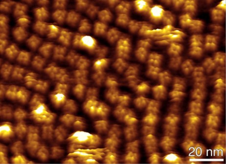
High-Resolution AFM Imaging
Membrane proteins are essential for life. They reside in lipidic membranes that form the boundaries of cells and their compartments. Membrane proteins are highly specialized molecular machineries that provide cellular membranes with unique functions. Cellular membranes peppered with membrane proteins not only separate chemically and physically different environments from each other but also actively create and maintain such differences. As such, they are involved in crucial processes such as energy conversion, signal transduction and amplification, enzymatic activities, molecular transport, anchoring of the cytoskeleton, formation of adhesion and motility. However, understanding how membrane proteins function requires to observe them at work.
Since its invention in 1986 by Gerber, Quate and Binnig, the atomic force microscope (AFM) has evolved into a multifunctional tool membrane protein research. In our research group we develop and employ these multifunctional tools to characterize membrane proteins, protein membranes and living cells. Here, however, we develop high-resolution AFM imaging methods approaching a lateral resolution of ≈1 nm and used them to gather information about the oligomeric state and assembly of membrane proteins and to observe membrane proteins at work.
Further reading
Quantifying and identifying two ligand-binding sites while imaging native human membrane receptors by AFM
M. Pfreundschuh, D. Alsteens, R. Wieneke, C. Zhang, S.R. Coughlin, R. Tampé, B.K. Kobilka & D.J. Müller
Nature Communications (2015) 6, 8857. external pageonline
Directly observing the lipid-dependent self-assembly and pore-forming mechanism of the cytolytic toxin listeriolysin O
E. Mulvihill, K. van Pee, S.A. Mari, D.J. Müller & Ö. Yildiz
Nano Letters (2015) 15, 6965-6973 external pageonline
Imaging G protein-coupled receptors while quantifying their ligand-binding free-energy landscape
D. Alsteens, M. Pfreundschuh, C. Zhang, P. Spoerri, S.R. Coughlin, B.K. Kobilka & D.J. Müller
Nature Methods (2015) 12, 845-851.
Localizing chemical groups while imaging single native proteins by high-resolution AFM
M. Pfreundschuh, D. Alsteens, M. Hilbert, M.O. Steinmetz & D.J. Müller
Nano Letters (2014) 14, 2957-2964.
Multiparametric high-resolution imaging of native proteins by force-distance curve– based AFM
M. Pfreundschuh, D. Martinez-Martin, E. Mulvihill, S. Wegmann & D.J. Muller
Nature Protocols (2014) 9, 1113-1130.
Quantitative imaging of the electrostatic field and potential generated by a transmembrane protein at sub-nanometer resolution
M. Pfreundschuh, U. Hensen & D.J. Muller
Nano Letters (2013) 13, 5585-5593.
Multi-parametric imaging of biological systems by force-distance curve-based AFM
Y.F. Dufrene, D. Martínez-Martín, I. Medalsy, D. Alsteens & D.J. Muller
Nature Methods (2013) 10, 847-854.
Nanomechanical properties of proteins and membranes depend on loading rate and electrostatic interactions
I. Medalsy & D.J. Muller
ACS NANO (2013) 7, 2642-2650.
High-resolution imaging of 2D outer membrane protein F crystals by atomic force microscopy
D. Fotiadis & D.J. Müller
Methods in Molecular Biology (2013) 736, 461-474.
Out but not in: β-strands shape the unfolding pathway but not the refolding of the large transmembrane β-barrel protein FhuA
J. Thoma, P. Bosshart, M. Pfreundschuh & D.J. Muller
Structure (2012) 20, 2185-2190.
Engineering rotor ring stoichiometries in ATP synthases
D. Pogoryelov, A.L. Klyszejko, G. Krasnoselska, E.M. Heller, V. Leone, J.D. Langer, J. Vonck, D.J. Muller, J.D. Faraldo-Gómez & T. Meier
Proc. Natl. Acad. Sci. USA (2012) 109, E1599-1608.
Investigating fibrillar aggregates of Tau protein by atomic force microscopy
S. Wegmann, D.J. Müller & E. Mandelkow
Methods in Molecular Biology (2012) 849, 169-183.
Gating of the MlotiK1 potassium channel involves large rearrangements of the cyclic nucleotide-binding domains
S.A. Mari, J. Pessoa, S.L. Altieri, U. Hensen, L. Thomas, J.H. Morais-Cabral & D.J. Muller
Proc. Natl. Acad. Sci. USA (2011) 108, 20802-20807.
Structure and function of the glucose PTS transporter from Escherichia coli
J.-M. Jeckelmann, D. Harder, S.A. Mari, M. Meury, Z. Ucurum, D.J. Muller, B. Erni & D. Fotiadis
Journal of Structural Biology (2011) 176, 395-403.
Quantifying chemical and physical properties of native membrane proteins at molecular resolution by force-volume AFM
I. Medalsy, U. Hensen & D.J. Muller
Angewandte Chemie International Edition (2011) 50,12103-12108.
High-resolution atomic force microscopy and spectroscopy of native membrane proteins
Ch. Bippes & D.J. Muller
Reports on Progress in Physics (2011) 74, 086601.
Studying collagen self-assembly by time-lapse high-resolution AFM
C. Franz & D.J. Müller
Methods in Molecular Biology (2011) 736, 97-107.
Human tau isoforms assemble into ribbon-like fibrils that display polymorphic structure and stability
S. Wegmann, Y.Y. Jung, S. Chinnathambi, E.M. Mandelkow, E. Mandelkow & D.J. Müller
Journal of Biological Chemistry (2010) 285, 27302-27313.
Assessing the structure and function of single biomolecules with scanning transmission electron and atomic force microscopes
S. Muller, D.J. Müller & A. Engel
Micron (2010) 42, 186-195.
pH induced conformational change of the outer membrane protein OmpG reconstituted into native E. coli lipids
S.A. Mari, C.A. Bippes, S. Köster, Ö. Yildiz, W. Kühlbrandt & D.J. Müller
Journal of Molecular Biology (2010) 396, 610-616.
The c13 ring from Bacillus TA2.A1 ATP synthase shows an extended diameter due to a special structural region
D. Matthies, L. Preiß, A.L. Klyszejko, D.J. Müller, G.M. Cook, J. Vonck & Th. Meier
Journal of Molecular Biology (2009) 388, 611-618.
Conformational adaptability of Redb during DNA annealing and implications for its structural relationship with Rad52
A. Erler, S. Wegmann, C. Elie-Caille, R. Bradshaw, M. Maresca, R. Seidel, B. Habermann, D.J. Müller & F. Stewart
Journal of Molecular Biology (2009) 391, 586-598.
AFM: A nanotool in membrane biology
D.J. Müller
Biochemistry (2008) 47, 7986-7998.
Vertebrate membrane proteins: Structure, function and insights from biophysical approaches
D.J. Müller, N. Wu & K. Palczewski
Pharmacological Reviews (2008) 60, 43-78.
Atomic force microscopy as a multifunctional molecular toolbox in nanobiotechnology
D.J. Müller & Y. Dufrene
Nature Nanotechnology (2008) 3, 261-269.
Strategies to prepare and characterize native protein membranes by AFM
D.J. Müller & A. Engel
Current Opinion in Colloid and Interface Science (2008) 13, 338-350.
An intermediate step in the evolution of ATPases - a hybrid FO-VO rotor in a bacterial Na+ F1FO ATP synthase
M. Fritz, A.L. Klyszejko, N. Morgner, J. Vonck, B. Brutschy, D.J. Müller, Th. Meier & V. Müller
FEBS Journal (2008) 275, 1999-2007.
Folding and assembly of proteorhodopsin
A.L. Klyszejko, S. Shastri, S.A. Mari, H. Grubmüller, D.J. Müller & C. Glaubitz
Journal of Molecular Biology (2008) 376, 35-41.
Straight GDP-tubulin protofilaments form in the presence of taxol
C. Elie-Caille, F. Severin, J. Helenius, J. Howard, D.J. Müller & A.A. Hyman
Current Biology (2007) 20, 1765-1770.
Atomic force microscopy and spectroscopy of native membrane proteins
D.J. Müller & A. Engel
Nature Protocols (2007) 2, 2191-2197.
The oligomeric state of c rings from cyanobacterial F-ATP synthases varies from 13 to 15
D. Pogoryelov, C. Reichen, A. Klyszejko, R. Brunisholz, D.J. Müller, P. Dimroth & Th. Meier
Journal of Bacteriology (2007) 189, 5895-5902.
Aminosulfonate modulated pH induced conformational changes in Connexin26 hemichannels
J. Yu, Ch. Bippes. G. Hand, D.J. Müller & G. Sosinsky,
Journal of Biological Chemistry (2007) 282, 8895-8904.
Imaging and detecting molecular interactions of single transmembrane proteins
H. Janovjak, A. Kedrov, D.A. Cisneros, K.T. Sapra, J. Struckmeier & D.J. Müller
Neurobiology of Aging (2006) 27, 546-561.
Structure of the rhodopsin dimer: A working model for G-protein coupled receptors
D. Fotiadis, B. Jastrzebska, A. Philippsen, D.J. Müller, K. Palczewski & A. Engel
Current Opinion in Structural Biology (2006) 16, 252-259.
A new approach to prepare membrane proteins for single molecule AFM imaging
D.A. Cisneros, D.J. Müller, S.M. Daud & J.H. Lakey
Angewandte Chemie International Edition (2006) 45, 3252-3256.
Structural evidence for a constant c11 ring stoichiometry in the sodium F-ATP synthase
Th. Meier, J. Yu, Th. Raschle, F. Henzen, P. Dimroth & D.J. Müller
FEBS Journal (2005) 272, 5474-5483.
The c15 ring of the Spirulina platensis F-ATP synthase: F1/Fo symmetry mismatch is not obligatory
D. Pogoryelov, J. Yu, Th. Meier, J. Vonck, P. Dimroth & D.J. Müller
Embo reports (2005) 6, 1040-1044.
Probing different origins of potential barriers stabilizing the membrane proteins halorhodopsin and bacteriorhodopsin
D. Cisneros, D. Oesterhelt & D.J. Müller
Structure (2005) 13, 235-242.
Atomic force microscopy of biological samples
P. Frederix, B.W. Hoogenboom, D. Fotiadis, D.J. Müller & A. Engel
MRS Bulletin (2004) 29, 449-455.
Observing membrane protein diffusion at subnanometer resolution
D. J. Müller, A. Engel, Th. Meier, U. Matthey, P. Dimroth & K. Suda
Journal of Molecular Biology (2003) 327, 925-930.
Observing structure function and assembly of single proteins by AFM
D.J. Müller, H. Janovjak, T. Lehto, L. Kuerschner & K. Anderson
Progress in Biophysics and Molecular Biology (2002) 71, 1-43.
Biomolecular imaging using atomic force microscopy
D.J. Müller & K. Anderson
Trends in Biotechnology (2002) 20, S45-S49.
Conformational changes in surface structures of isolated Connexin26 gap junctions
D.J. Müller, G.M. Hand, A. Engel & G.E. Sosinsky
EMBO Journal (2002) 21, 3598-3607.
Identification and structure of a putative Ca2+-binding domain at the C terminus of AQP1
D. Fotiadis, K. Suda, P. Tittmann, P. Jenö, A. Philippsen, D.J. Müller, H. Gross & A. Engel
Journal of Molecular Biology (2002) 318, 1381-1394.
Imaging the electrostatic potential of transmembrane channels: Atomic probe microscopy of OmpF porin
A. Philippsen, W. Im, A. Engel, T. Schirmer, B. Roux & D.J. Müller
Biophysical Journal (2002) 82, 1667-1676.
Sampling the conformational space of membrane protein surfaces with the AFM
S. Scheuring, D.J. Müller, H. Stahlberg, H.-A. Engel & A. Engel
European Biophysical Journal (2002) 31, 172-178.
The central plug in the reconstituted undecameric c cylinder of a bacterial ATP synthase consists of phospholipids
Th. Meier, U. Matthey, F. Henzen, P. Dimroth, & D.J. Müller
FEBS Letters (2001) 505, 353-356.
Subunits constrain stoichiometry of ATP synthase rotor
D.J. Müller, N. Dencher, Th. Meier, P. Dimroth, H. Stahlberg, K. Suda, A. Engel, H. Seelert & U. Matthey
FEBS Letters (2001) 504, 219-222.
Bacterial sodium ATP synthase has an undecameric motor
H. Stahlberg, D.J. Müller, K. Suda, D. Fotiadis, A. Engel, Th. Meier, U. Matthey & P. Dimroth
EMBO Reports (2001) 2, 229-233.
From images to interactions: High-resolution phase imaging in tapping-mode atomic force microscopy
M. Stark, C. Möller, D.J. Müller & R. Guckenberger
Biophysical Journal (2001) 80, 3009-3018.
Observing proteins at work with the atomic force microscope
A. Engel & D.J. Müller
Nature Structural Biology (2000) 7, 715-718.
Reversible loss of crystallinity during photobleaching purple membrane
C. Möller, G. Büldt, N.A. Dencher, A. Engel & D.J. Müller
Journal of Molecular Biology (2000) 301, 869-879.
The surface topography of lens MIP supports dual functions
D. Fotiadis, L. Hasler, D.J. Müller, H. Stahlberg, J. Kistler & A. Engel
Journal of Molecular Biology (2000) 300, 779-789.
Atomic force microscopy of native purple membrane
D.J. Müller, J.B. Heymann, F. Oesterhelt, C. Möller, H. Gaub, G. Büldt & A. Engel
Biochemical Biophysical Acta (2000) 1460, 27-38.
Conformational changes, flexibilities and intramolecular forces observed on individual proteins using AFM
D.J. Müller, D. Fotiadis, C. Möller, S. Scheuring & A. Engel
Single Molecule (2000) 1, 115-118.
Conformations of the rhodopsin third cytoplasmic loop grafted onto bacteriorhodopsin
J.B. Heymann, M. Pfeiffer, V. Hildebrandt, D. Fotiadis, B. de Groot, R. Kabak, A. Engel, D. Oesterhelt & D.J. Müller
Structure (2000) 8, 643-653.
Proton powered turbine of a plant motor
H. Seelert, A. Poetsch, N. Dencher, A. Engel, H. Stahlberg & D.J. Müller
Nature (2000) 405, 418-419.
Unfolding pathways of individual bacteriorhodopsins
F. Oesterhelt, D. Oesterhelt, M. Pfeiffer, A. Engel, H. Gaub & D.J. Müller
Science (2000) 288, 143-146.
Atomic force microscopy: a powerful tool to observe biomolecules at work
A. Engel, Y. Lyubchenko & D.J. Müller
Trends in Cell Biology (1999) 9, 77-80.
Atomic force microscopy: a forceful way with single molecules
A. Engel, H. Gaub & D.J. Müller
Current Biology (1999) 9, R133-R136.
Charting the surfaces of purple membrane
J.B. Heymann, D.J. Müller, E. Landau, J. Rosenbusch, E. Pebay-Peroulla, G. Büldt & A. Engel
Journal of Structural Biology (1999) 128, 243-249.
Controlled unzipping of a bacterial surface layer with atomic force microscopy
D.J. Müller, W. Baumeister & A. Engel
Proc. Natl. Acad. Sci. USA (1999) 96, 13170-13174.
High resolution topographs of the Escherichia coli waterchannel aquaporin Z
S. Scheuring, P. Ringler, M. Borgina, H. Stahlberg, D.J. Müller, P. Agre & A. Engel
EMBO Journal (1999) 18, 4981-4987.
Tapping mode atomic force microscopy produces faithful high-resolution images of protein surfaces
C. Möller, M. Allen, V. Elings, A. Engel & D.J. Müller
Biophysical Journal (1999) 77, 1150-1158.
Surface structures of native bacteriorhodopsin depend on the molecular packing arrangement in the membrane
D.J. Müller, H.-J. Sass, S.A. Müller, G. Büldt & A. Engel
Journal of Molecular Biology (1999) 285, 1903-1909.
pH and voltage induced structural changes of porin OmpF explains channel closure
D.J. Müller & A. Engel
Journal of Molecular Biology (1999) 285, 1347-1351.
Electrostatically balanced subnanometer imaging of biological specimens by atomic force microscope
D.J. Müller, D. Fotiadis, S. Scheuring, S.A. Müller & A. Engel
Biophysical Journal (1999) 76, 1101-1111.
Imaging streptavidin 2D crystals on biotinylated lipid monolayers at high resolution with the atomic force microscope
S. Scheuring, D.J. Müller, P. Ringler, B. Heyman & A. Engel
Journal of Microscopy (1999) 193, 28-33.
Surface analysis of the photosystem I complex by electron and atomic force microscopy
D. Fotiadis, D.J. Müller, G. Tsiotis, L. Hasler, P. Tittmann, T. Mini, P. Jenö, H. Gross & A. Engel
Journal of Molecular Biology (1998) 283, 83-94.
Mapping flexible protein domains at subnanometer resolution with the AFM
D.J. Müller, D. Fotiadis & A. Engel
FEBS Letters (1998) 430, 105-111.
High-resolution imaging of native biological sample surfaces with scanning probe microscope
A. Engel, C.-A. Schoenenberger & D.J. Müller
Current Opinion in Structural Biology (1997) 7, 279-284.
Electron and atomic force microscopy of membrane proteins
J.B. Heymann, D.J. Müller, K. Mitsuoka & A. Engel
Current Opinion in Structural Biology (1997) 7, 543-549.
Structural changes of native membrane proteins monitored at subnanometer resolution with the atomic force microscope
D.J. Müller, C.-A. Schoenenberger, F. Schabert & A. Engel
Journal of Structural Biology (1997) 119, 149-157.
The height of biomolecules measured with the atomic force microscope depends on electrostatic interactions
D.J. Müller & A. Engel
Biophysical Journal (1997) 73,1633-1644.
Adsorption of biological molecules to a solid support for scanning probe microscopy
D.J. Müller, M. Amrein & A. Engel
Journal of Structural Biology (1997) 119, 172-188.
The bacteriophage ø29 head-tail connector imaged at high resolution with atomic force microscopy
D.J. Müller, A. Engel, J.L.Carrascosa & M. Velez
EMBO Journal (1997) 16, 101-107.
Immuno atomic force microscopy of purple membranes
D.J. Müller, C.-A. Schoenenberger, G. Büldt & A. Engel
Biophysical Journal (1996) 68, 1681-1686.
Conformational change of the hexagonally packed intermediate layer of Deinococcus radiodurans observed with atomic force microscopy
D.J. Müller, W. Baumeister & A. Engel
Journal of Bacteriology (1996) 178, 3325-3329.
Surface topographies at subnanometer-resolution reveal asymmetry and sidedness of aquaporin-1
T. Walz, P. Tittmann, K.H. Fuchs, D.J. Müller, B.L. Smith, P. Agree, H. Gross & A. Engel
Journal of Molecular Biology (1996) 264, 907-918.
Force-induced conformational change of bacteriorhodopsin
D.J. Müller, G. Büldt & A. Engel
Journal of Molecular Biology (1995) 249, 239-243.
Imaging purple membranes in aqueous solutions at sub-nanometer resolution by atomic force microscopy
D.J. Müller, F.A. Schabert, G. Büldt & A. Engel
Biophysical Journal (1995) 68, 1681-1686.