Developing Biophysical Tools

Our group is interested in understanding how membrane proteins fold, how they work in vitro, how they work in vivo, and how membrane proteins collectively contribute to cellular functions and processes including homeostasis, cell shape, adhesion and communication. To contribute to this understanding we develop mostly AFM-based nanotechnological tools that allow to image single native membrane proteins in vitro and in vivo at sub-nanometer resolution. Other AFM-based tools allow to directly observe the folding steps of single membrane proteins in the native membrane or to quantify and localize interactions stabilizing membrane proteins. Again other methods allow to characterize the contribution of individual membrane proteins to cellular processes including cell adhesion or the mechanisms guiding the drastic cell shape changes in mitosis. In the following we describe some of these tools, methods and approaches and exemplify how we apply these to address pertinent questions in life sciences. For detailled reading we list the scientific publications our reserach group has published. We also kindly refeer to the link 'Adressing Biological Problems'.
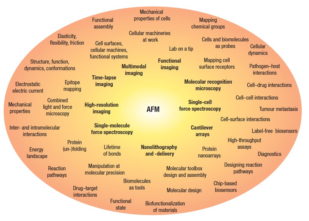
Atomic force microscopy (AFM)-based approaches, which have been developed to characterize and manipulate biological systems on the nanoscale. Originally invented to image the topography of surfaces (Binnig et al. Phys. Rev. Lett (1986) 56, 930-933), the AFM has evolved into a multifunctional nanotool that makes it possible to characterize tissues, cells, membranes, proteins and DNA/RNA with unprecedented resolution. AFM-based methods allow single-molecule analyses and measuring of various biochemical, physical and chemical properties and interactions of living systems. Figure taken from Muller & Dufrene, Nature Nanotechnology (2008) 3, 261-269.
Atomic force microscopy (AFM)-based approaches, which have been developed to characterize and manipulate biological systems on the nanoscale. Originally invented to image the topography of surfaces (Binnig et al. Phys. Rev. Lett (1986) 56, 930-933), the AFM has evolved into a multifunctional nanotool that makes it possible to characterize tissues, cells, membranes, proteins and DNA/RNA with unprecedented resolution. AFM-based methods allow single-molecule analyses and measuring of various biochemical, physical and chemical properties and interactions of living systems. Figure taken from Muller & Dufrene, Nature Nanotechnology (2008) 3, 261-269.
JavaScript has been disabled in your browser